 Home
Home
Knee Ligament Reconstruction
Restoring Strength and Stability to Your Knee
Understanding Knee Anatomy
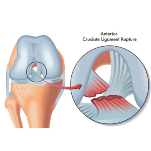
The knee is one of the most complex and essential joints in the human body. It connects the thighbone (femur) and the shinbone (tibia), with the kneecap (patella) sitting at the front to provide additional protection. Stability and movement are controlled by four strong ligaments, which act like ropes to keep the joint in place: 🔹 Collateral Ligaments – Located on the inner and outer sides of the knee, they control side-to-side movements. 🔹 Cruciate Ligaments (ACL & PCL) – Found at the center of the knee joint, these ligaments cross to form an "X" shape. They control back-and-forth motion and rotational stability.
What Causes Knee Ligament Injuries?
Knee ligament injuries are common in sports that involve sudden stops, changes in direction, and pivoting, such as: ⚽ Football 🏀 Basketball 🏒 Hockey 🏉 Rugby Ligament injuries are classified into three grades based on severity: ✔️ Grade I – Mild stretching of the ligament with no significant instability. ✔️ Grade II – Partial tear of the ligament, causing moderate instability. ✔️ Grade III – Complete ligament rupture, leading to joint instability and loss of function.
What is Arthroscopic Knee Ligament Reconstruction?
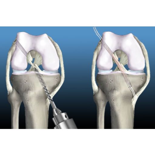
Ligament reconstruction is a minimally invasive procedure designed to restore knee stability by replacing the damaged ligament with a tissue graft. The graft can be taken from: 🦵 Your own body (autograft) – Typically from the hamstring, patellar tendon, or quadriceps tendon. 🔬 A donor (allograft) – Used in certain cases for faster recovery and reduced surgical trauma. The new ligament is secured to the femur and tibia using specialized screws, clips, or staples. Over time, the bone integrates with the graft, making it a natural part of the knee.
How is Arthroscopic Knee Ligament Reconstruction Performed?
This advanced surgical technique uses a tiny camera (arthroscope) and specialized instruments, inserted through small incisions around the knee. The procedure follows these steps: 1️⃣ Anesthesia – General or regional anesthesia is administered for patient comfort. 2️⃣ Joint Visualization – A sterile solution is introduced to expand the knee joint, allowing for a clear view. 3️⃣ Arthroscope Insertion – The surgeon examines the knee joint on a high-definition monitor. 4️⃣ Damaged Ligament Removal – Any remaining torn ligament is carefully removed. 5️⃣ Graft Placement – A new ligament is positioned and fixed securely to the femur and tibia. 6️⃣ Closure – The incisions are closed with sutures or adhesive strips, and a sterile bandage is applied.
Benefits of Arthroscopic Knee Ligament Reconstruction
Unlike traditional open surgery, arthroscopic ligament reconstruction offers multiple advantages: ✔️ Minimally invasive – Smaller incisions mean less trauma to surrounding tissues. ✔️ Reduced postoperative pain – Faster pain relief and quicker return to daily activities. ✔️ Shorter hospital stay – Most patients can go home the same day. ✔️ Faster recovery – Less downtime and quicker return to sports.
Recovery & Rehabilitation
While knee ligament reconstruction restores stability, rehabilitation is key to regaining full function. 📅 First Weeks: Focus on pain management, swelling control, and gentle mobility exercises. 🏋️ Months 2-3: Progressive strengthening of the knee and surrounding muscles. 🏃 Months 4-6: Sport-specific training to restore agility, balance, and endurance. 🥇 Return to Play: Most athletes can safely resume high-impact sports after 6 to 12 months, depending on progress.
Regain Confidence in Your Knee
A torn ligament doesn’t have to mean the end of an active lifestyle. With expert surgical care and a personalized rehabilitation program, you can return to the activities you love—stronger than ever.